A cranioplasty describes the replacement of native cranial-bone in order to close an existing defect for functional and aesthetic restoration.
In the last 400 years, several techniques and materials have been applied for cranial defect closure. Even before that, as an unverified, ancient skull containing a metal implant featured in the Museum for Osteology in Oklahoma, suggests that cranioplasty has been a practiced procedure thousands of years ago using metals.
In ancient and medieval times, trepanations, so creating a burr hole in the skull, used to be a common practice after e. g. heavy trauma due to weapon impact, accidents or animal attacks. It was used to allow rearrangement of cranial bone fragments and release of blood-buildup below the cranium after trauma, but it was also believed to release evil spirits that were associated with abnormal or insane behaviors. There is a surprisingly low putative mortality rate for trepanations of below 10 %, indicating a low infection rate in the absence of antibiotics or sterility precautions.
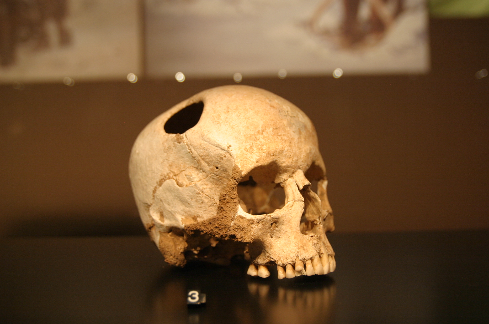
Picture source: Rama on wikipedia/CC BY-SA 3.0 FR
However, these procedures left permanent defects, leaving the brain vulnerable. Reports about the previously mentioned ancient Peruvian society indicate that coconut shells or even palm leaves were used as cover and suggest that choice of material was status-dependent.
The first report and description of cranioplasty in Europe was made by Gabriele Fallopius in the 16th century, stating that the fractured cranium should be removed and be reinserted with a gold plate if the dura was damaged.
The first documented procedure of crafting a cranioplastic using canine-skull bone was performed by the Dutch surgeon Job van Meekeren in 1668 on a Russian citizen. Threatened to be excommunicated, the patient had to leave Russia due to the Church’s outrage about the procedure.
In general, for defect closure, several material-options are available. The patient’s native bone (autografts), animal bone (allografts), organic polymers, metals or ceramics (alloplasts), each with their individual advantages and properties. While native bone is favored due to its biocompatibility, infection resistance, stiffness and elastic properties, the primary disadvantage of native bone is its availability. Bone for closure of cranial defects needs to be harvested from other regions in the patient, for example the shoulder or hip. However, when harvesting autologous bone for cranial defect closure, this automatically requires a secondary surgery, which carries further risks like donor-site morbidity, donor-site pain, additional anesthesia or lack of shaping options. Aside the fact that there is only a finite amount of native bone harvestable, there is always a risk of autograft resorption due to immunological rejection.
Alternatively, in former times, animal bone was used but carries elevated risks of transferable diseases and immunorejection. This option was practiced before but was especially common in WWI due to resource-scarce conditions and the destructive nature of 20th century warfare.
Today, these kinds of defects on the cranium stem from craniectomies, trepanations, traumatic head injuries, infections or oncological resections. The created defects can impair the patient functionally and aesthetically, reducing confidence in their appearance, which in turn affects their quality of life significantly. Therefore, a practically unlimited amount of available alloplast materials nowadays, ensures a broad range of patient care. Each material has its specific properties and thus advantages. What materials exactly are used today and why? How and why did practice evolve in this direction? We will have a look at these questions in the next article. [03/2026 NK]
Quellen
S. Honeybul, K. M. Ho: Cranioplasty: morbidity and failure. In: British journal of neurosurgery, May 2016, doi:10.1080/02688697.2016.1187259, PMID 27215939.
C. B. Courville: Cranioplasty in prehistoric times. In: Bulletin of the Los Angeles Neurological Society, March 1959, Band 24, Nr. 1, S. 1–8, PMID 13629188.
Sanan, S. J. Haines: Repairing holes in the head: a history of cranioplasty. In: Neurosurgery, March 1997, Band 40, Nr. 3, S. 588–603, PMID 9055300.
Dujovny M, Aviles A, Agner C, Fernandez P, Charbel FT: “Cranioplasty: cosmetic or therapeutic?”. In: Surgical Neurology, March 1997, 47 (3): 238–41, https://doi.org/10.1016/S0090-3019(96)00013–4
Najeeb S, Zafar MS, Khurshid Z, Siddiqui F: Applications of polyetheretherketone (PEEK) in oral implantology and prosthodontics. In: J Prosthodont Res., January 2016, 60(1):12–9, doi: 10.1016/j.jpor.2015.10.001, PMID: 26520679