
As already mentioned in the history of cranioplasty, thousands of years ago, the first primitive cranioplasty procedures used, among other things, precious metals; however, in the late 19th century, the first bone grafts were used to repair cranial defects. The two world wars of the 20th century and the numerous associated injuries demonstrated that usage of autologous bone was insufficient due to its limited availability, coupled with increased infection rates and the need for a secondary operation at the patient’s bone donor site.
Matsuno et al. report infection rates of over 25 % with the use of autologous bone. In this context, experiments were also conducted using animal bones from dogs, monkeys, geese, rabbits, or calves. In 1901, Marchand reported that boiled and perforated animal horns—such as those from buffalo or even ivory were particularly biocompatible.
In 1915, Morestin reported the use of human cadaver cartilage to cover a cranial defect. However, this cartilage showed no significant calcification at the implantation site and was generally too weak to provide adequate mechanical protection. Sterilization and preparation of the material for implantation further weakened it. Due to high rates of infection and resorption, this treatment method did not gain acceptance in the long term.
This period also saw attempts to use strong, malleable and more easily sterilizable materials such as aluminum. Unfortunately, this material caused inflammation of the surrounding soft tissue, seizures, and slowly dissolved at the implantation site, which ultimately led to the abandonment of this material. Other metals such as lead, tantalum, or even platinum were used. While lead resulted in predictable signs of toxicity and related fatalities, tantalum and platinum were used with some success; however, due to their high material cost, they were not suitable for mass production.
Platinum in particular, demonstrated good biocompatibility and no signs of inflammation in the surrounding tissue. The situation was similar with tantalum, whose use, however, resulted in cases of headaches caused by sensitivity to weather changes, due to the material’s high thermal conductivity. Gold and silver also yielded similarly positive results as platinum but shared the same disadvantage of high cost. Further, silver caused discoloration of the scalp.
In addition to metals, autografts, allografts, and polymers, attempts were also made using inorganic ceramics to find a suitable bone substitute. It makes sense to replace natural bone with calcium phosphate, more specifically with hydroxyapatite (Ca5[OH|(PO4)3]) the native structural component of bone. Advantages of this material include good biocompatibility and osseointegration, as well as good vascularization due to the porosity of the material. Disadvantages include pronounced brittleness and fragility, as with many ceramics, as well as the potential for bacteria to adhere within the porous material. Nevertheless, for many years, hydroxyapatite was the preferred material for closing cranial hard tissue defects, although good osseointegration in patients is often not achieved until after 6 months or more. In the meantime, wearing a helmet can protect against fracture of the implant.
Nowadays, polymethyl methacrylate (PMMA) is often used to cover cranial defects; it was first used in patients in the 1950s. The liquid methacrylate monomer is polymerized with or without a mold, using a radical initiator under conditions of intense heat generation in the operating room, resulting in hardened (and shaped) PMMA that can be cut to size. With this method of production, however, not all radicals and monomers can ever be washed out of the process, so residual molecules always remain and are released at the implantation site. This can lead to inflammation or irritation of the surrounding tissue, and in severe cases may necessitate explantation. In in vivo applications of PMMA, such as total hip arthroplasty, the implant is fabricated directly within the joint at the patient’s implantation site. With this procedure, even larger amounts of radicals and MMA monomers are released into the patient, which can lead to a bone-cement reaction, with—depending on the severity—reported 30-day mortality rates of up to 88 %.
In addition to PMMA, there is a whole range of other materials used in the the manufacture of patient-specific skull implants. Each of these materials has its own advantages and disadvantages, and at present, it is not possible to recommend a single “best” material across the board for every application and every patient. The choice of the material best suited to the defect is at the discretion of the treating physician and should be carefully considered and evaluated on a case-by-case basis.
Based on the experiences and findings mentioned above, ideal cranioplasty materials should have the following properties:
- Adapts perfectly to the defect and anatomy
- Radiolucency for improved (post-operative) diagnostics
- Resistant to infection, with biofilm- and germ-repellent surface properties
- No shrinkage or expansion when heated
- Fracture-resistant yet elastic
- Bone-like properties
- Easy to work with intraoperatively
- Cost-effective
- Easy and quick to use
At 3di, we specialize in three materials that meet the high standards for a bone substitute material in the cranial region and have been proven over multiple decades. This allows us to respond flexibly to various medical requirements and offer solutions for a wide range of defects. The implant materials we use are the antimicrobial glass-ceramic BIOVERIT®II, the metallic titanium alloy Ti6Al4V, and the thermoplastic polyetheretherketone (PEEK). Our patient-specific implants are currently manufactured using CNC milling right here in Jena, eliminating time-expenses and other risks associated with on-site fabrication in the operating room.
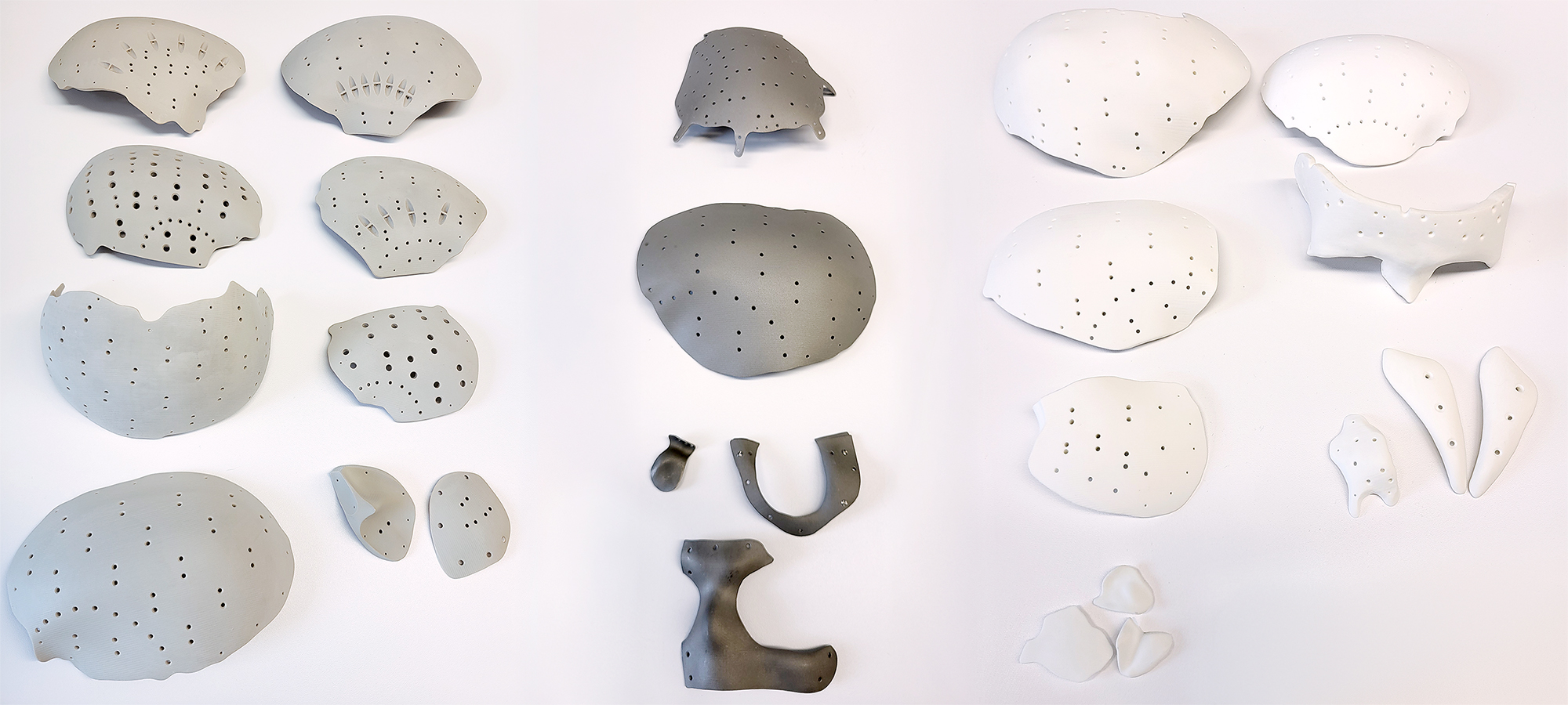
Picture source: 3di GmbH (CD)
We will evaluate these three materials we are using in more detail in upcoming posts and present even more interesting facts.
By the way: Our prices are independent of the material used. The decision on the appropriate material is based on the specific defect and tailored individually to each patient.
Quellen
Durand JL, Renier D, & Marchac D: The history of cranioplasty. Ann Chir Plast Esthet 42:75–83, 1997. (FRA)
Matsuno A, Tanaka H, Iwamuro H, Takanashi S, Miyawaki S, & Nakashima M, et al.: Analyses of the factors influencing bone graft infection after delayed cranioplasty. Acta Neurochir (Wien) 148:535–540, 2006
Black SP: Reconstruction of the supraorbital ridge using aluminum. Surg Neurol. 1978;9:121–8. PMID:625697
Siracusa V, Maimone G, Antonelli V. State-of-Art of Standard and Innovative Materials Used in Cranioplasty. Polymers (Basel). 2021 Apr 30;13(9):1452. doi: 10.3390/polym13091452, PMID:33946170, PMCID: PMC8124570.
Gaik C, Schmitt N, Wiesmann T: Knochenzementreaktion – Pathophysiologie, Diagnostik und Behandlungsoptionen. Anästh Intensivmed 2019;60:124–133. DOI: 10.19224/ai2019.124
Grant GA, Jolley M, Ellenbogen RG, Roberts TS, Gruss JR, & Loeser JD: Failure of autologous bone-assisted cranioplasty following decompressive craniectomy in children and adolescents. J Neurosurg 100:2 Suppl Pediatrics 163–168, 2004
Kühn K‑D, Lieb E, Berberich C (2016:) PMMA bone cement: what is the role of local antibotics? Maîtrise orthopédique, Proceeding of N°243, commission paritaire 1218T86410, S 12–18 (1148 2362)
Aydin S, Kucukyuruk B, Abuzayed B, Aydin S, Sanus GZ. Cranioplasty: Review of materials and techniques. J Neurosci Rural Pract. 2011 Jul;2(2):162–7. doi: 10.4103/0976–3147.83584, PMID: 21897681, PMCID: PMC3159354.