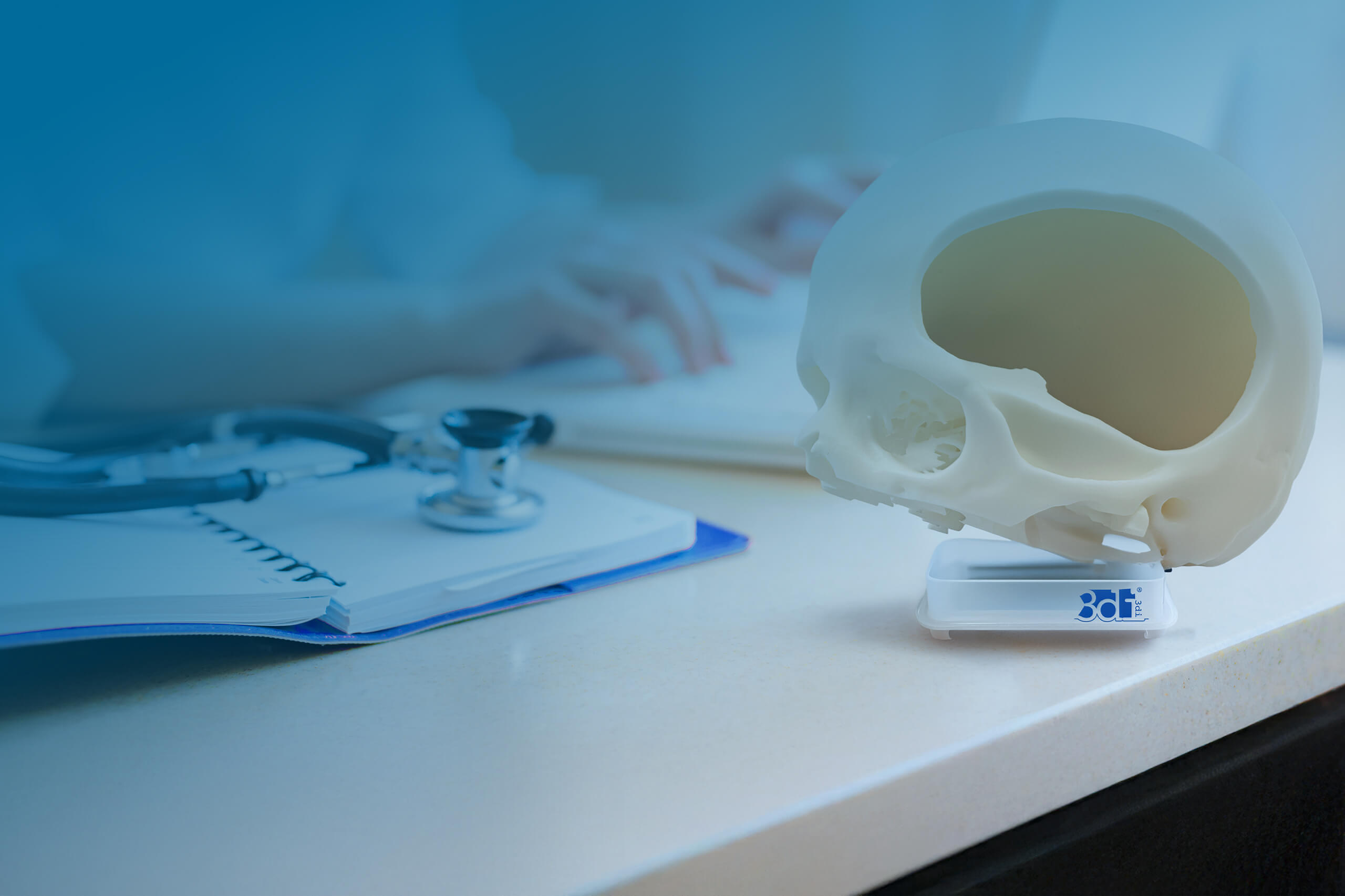
Hard tissue models
All about
hard tissue models
Our hard tissue models are used for consultation and surgical planning, as object for demonstration purposes, for training and education, and in research and development. With the help of real CT data, detailed, patient-specific models can also be created.
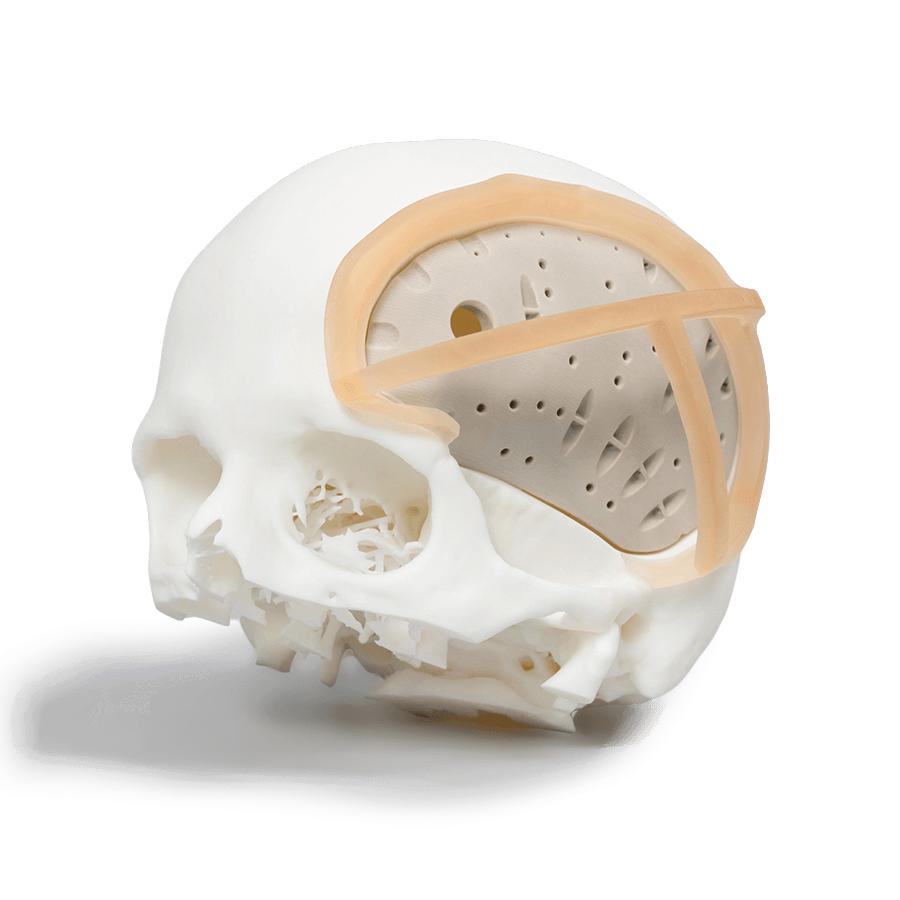
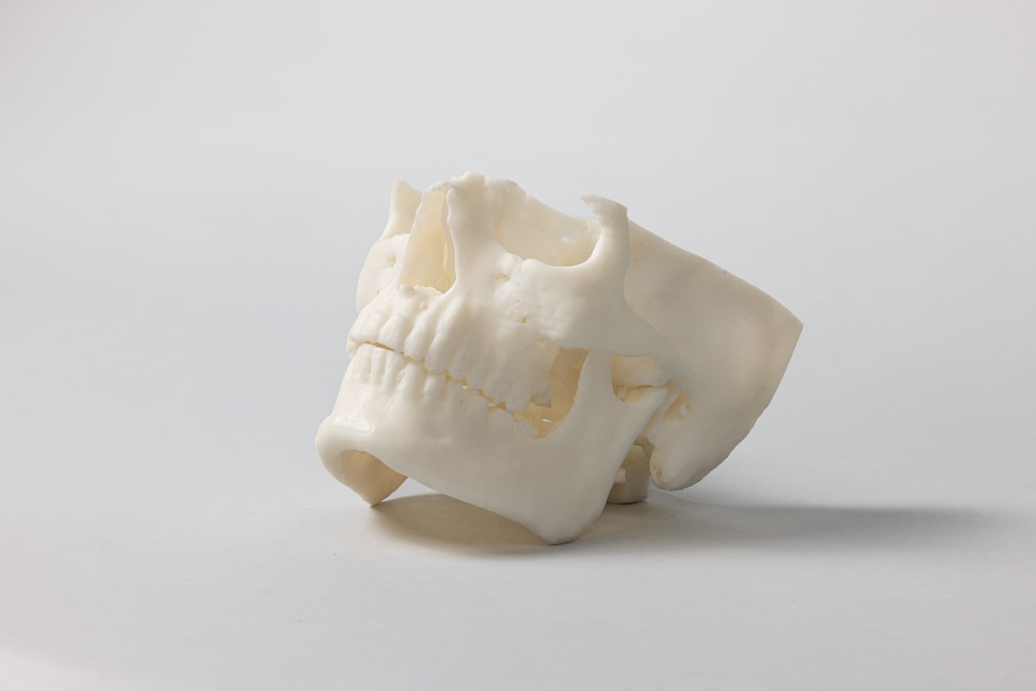
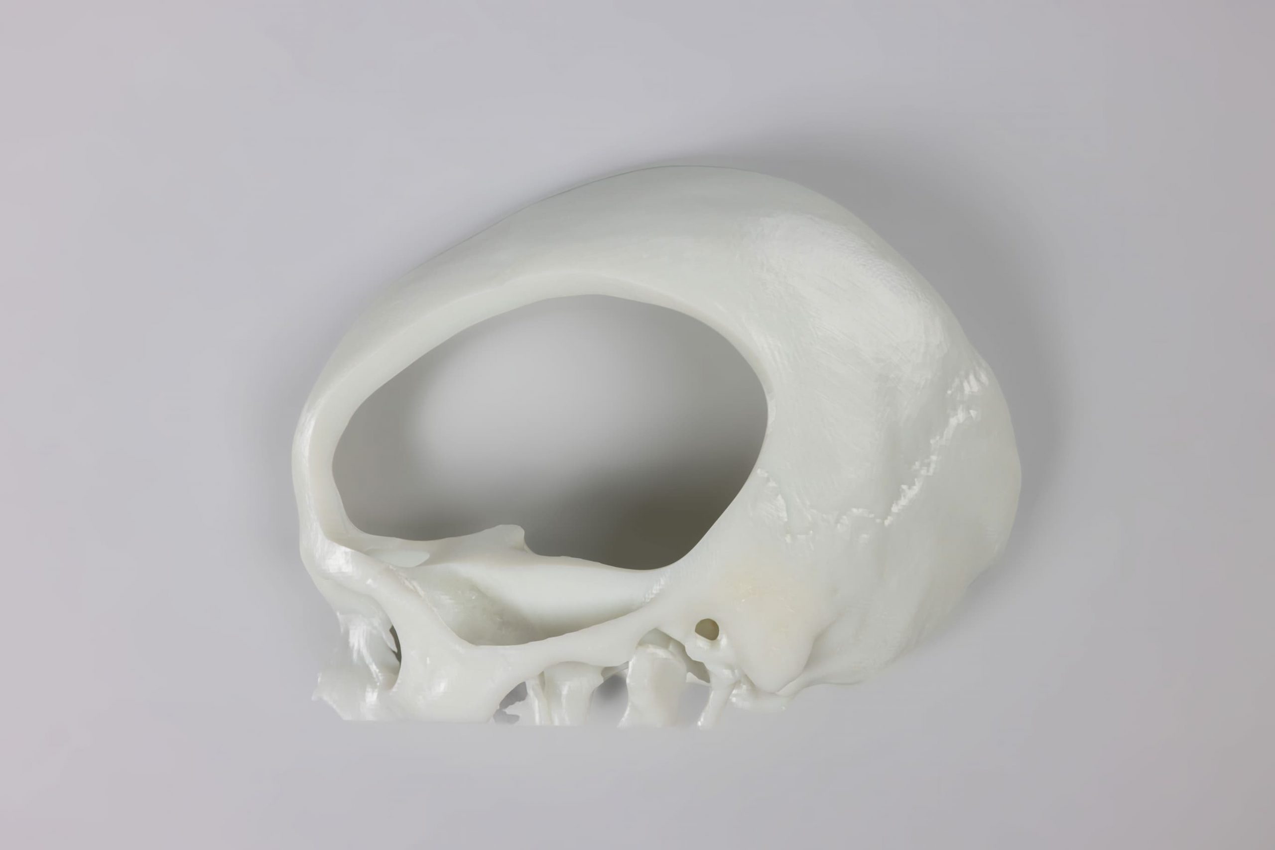
Skull models
In complex cases, even more detailed surgical planning can be helpful and useful. Upon request, we can produce models of the patient’s skull (or skull section) and the implant, as well as templates if necessary. The skull models can be edited by the physician and thus provide important information for planning the actual implant.
Material
Acrylate-based synthetic resin (methacrylate), steam sterilisable, suitable for short-term use on patients (e. g. for resection templates).
Advantages
- Detailed representation of bone structures
- Excellent workability for realistic surgical planning (non-sterile)
- Lower costs due to upstream, more accurate surgical planning
- Rapid availability
Application
- Surgical planning
- Research and development
- Training and advanced education
- Demonstration objects
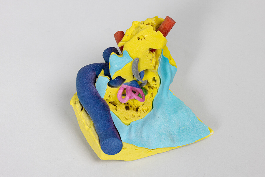
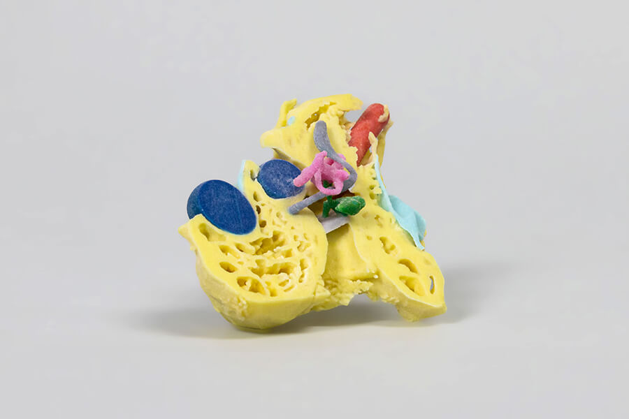
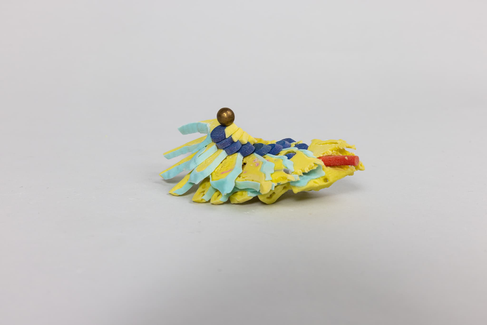
Petrous bone models
The complex anatomy of the petrous bone demands extensive training before microsurgery should be performed on the middle ear. For this training, we provide a three-dimensional petrous bone model, developed in collaboration between the Department of Otorhinolaryngology at the University of Jena and 3di GmbH.
Petrous bone model for preparation
Advantages
- Anatomical structures are easy to distinguish thanks to coloured backgrounds
- Simplified learning during dissection
Application
- Specialist training
- General medical training and continuing education
- Illustrative objects
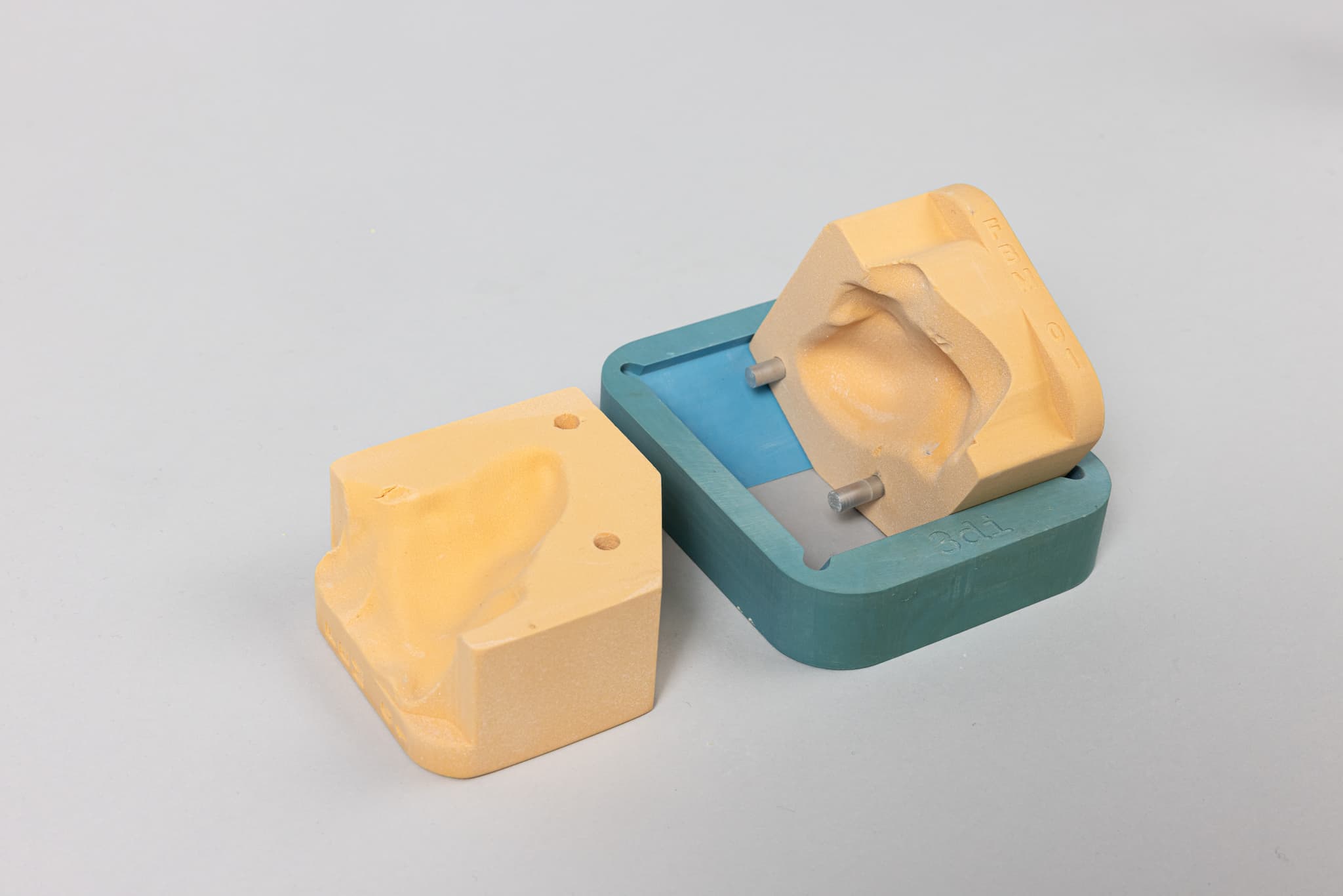
Additional option:
mounting device for petrous bone model
The holder is an ideal addition and facilitates training on the petrous bone model.
Advantages
- Simulation of the operation in a natural position
- The model cannot slip and does not need to be secured in any other way
- Hands can be placed comfortably
- The holder is reusable
Petrous bone models for illustration purposes
Representation and illustration of the positional relationships of the internal structures of the human petrous bone.
Axial section, sagittal section, open version, halved, open version with bridge
Application
- Specialist training
- General medical training and advanced education
- Illustrative objects (mastoidectomy)
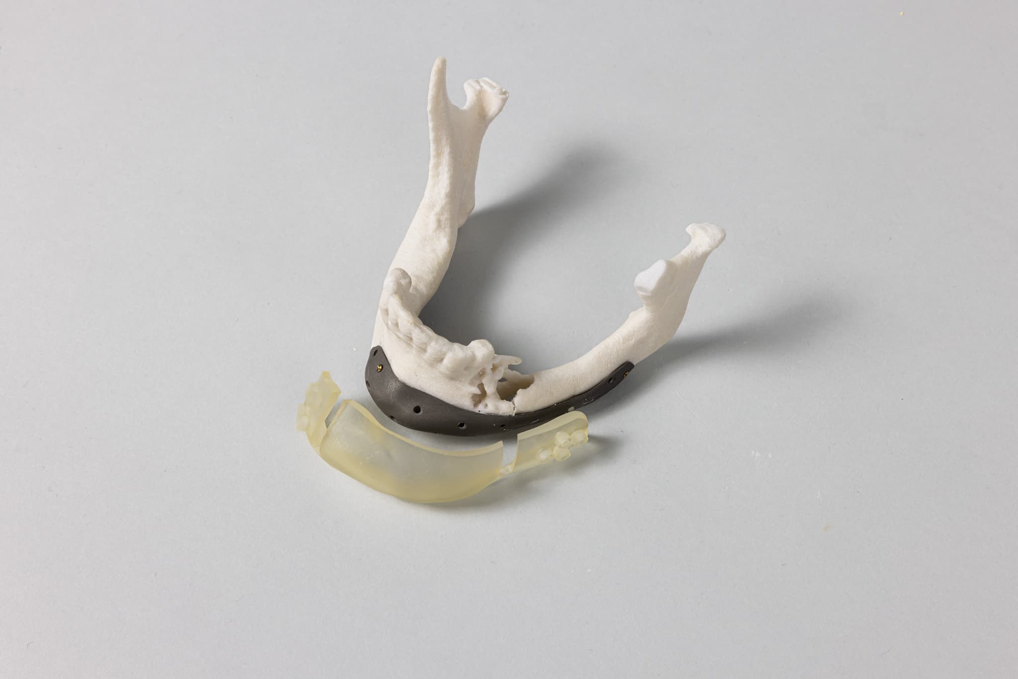
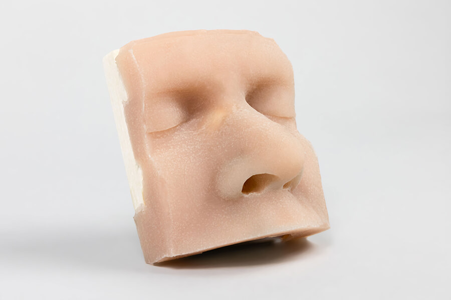
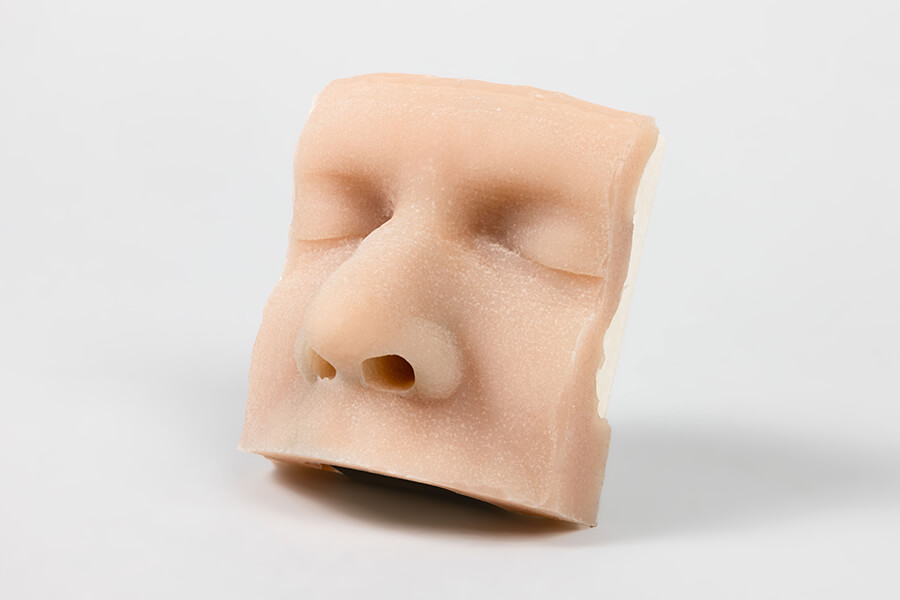
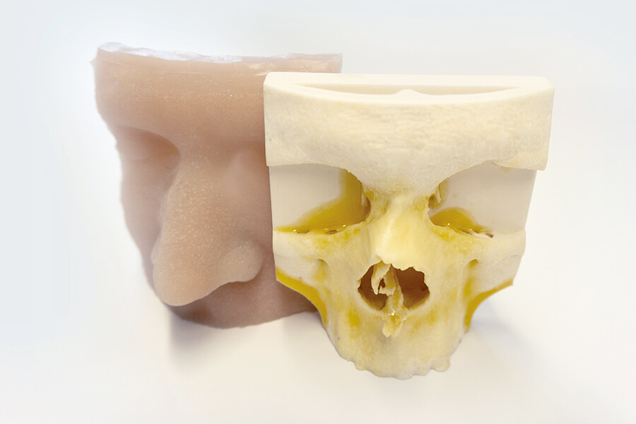
Osteotomy models
In collaboration with the ENT University Clinic in Jena and the company 3di GmbH, a training model for operations on the bony bridge of the nose was developed. The bony part of the model is covered with a removable silicone layer to make the surgical simulation as realistic as possible.
Advantages
- Surgery simulation on the model for training various skills, such as:
- Vestibulum margin incision, tunnelling and identification of the aperture
- Palpation of the aperture edges
- Intercartilaginous incision, tunnelling of the nasal bridge
- Palpation of the bony nasal bridge
- Paramedian osteotomies on both sides
- Lateral osteotomies on both sides
- Transverse osteotomies on both sides
Application
- Specialist training
- General medical training and advanced education
- Illustrative objects
Postcranial models
By the way: using real CT/MRI data, we can also produce detailed postcranial models for you according to your specifications. Please contact us!
Material
Acrylate-based synthetic resin (methacrylate), steam sterilisable
Advantages
- Detailed representation of bone structures
- Excellent workability for realistic surgical planning (non-sterile)
- Lower costs due to upstream, more accurate surgical planning
Application
- Surgical planning
- Research and development
- Training and advanced education
- Illustrative objects
Do you have any further questions?


