Soft tissue models
All about
soft tissue models
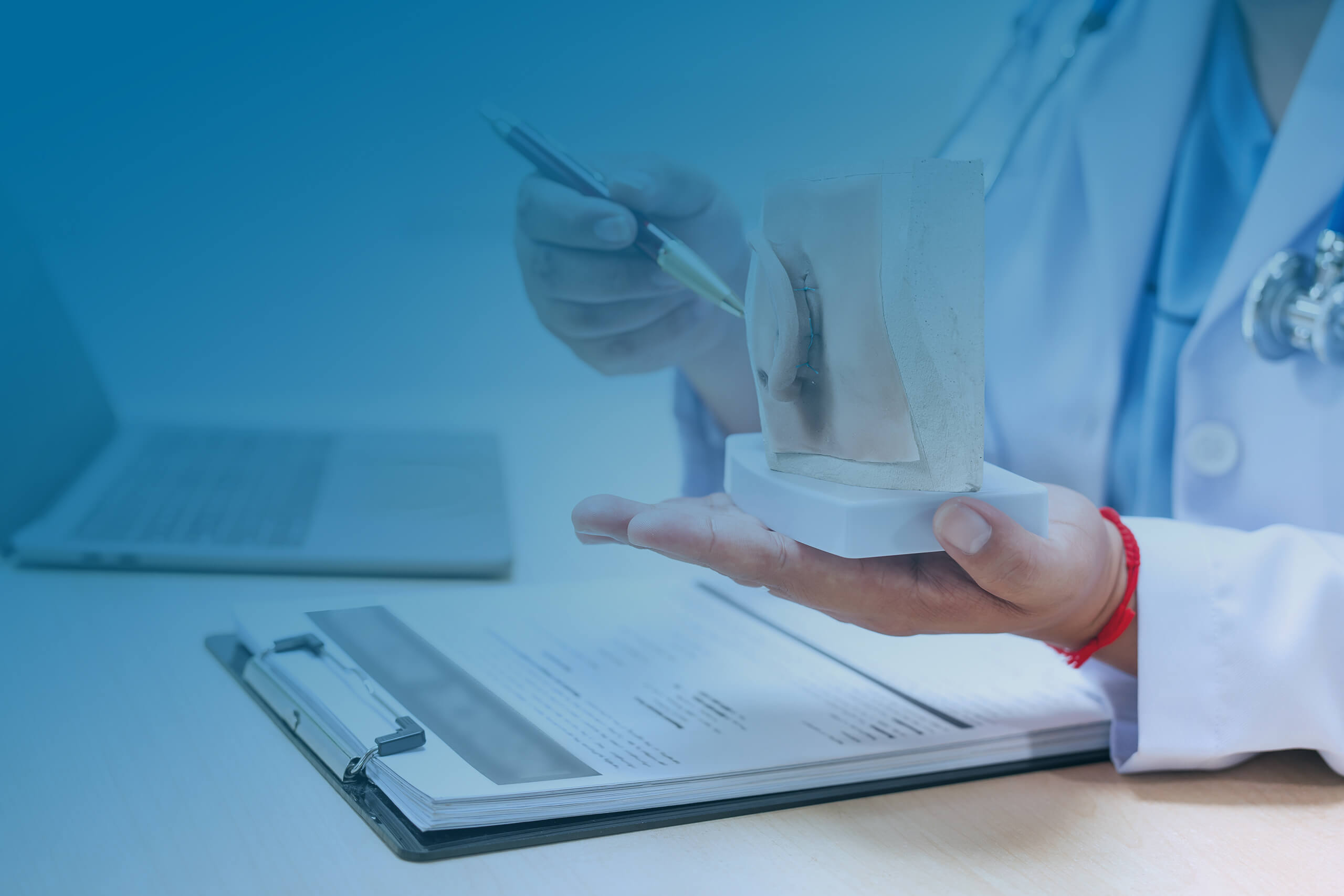
In collaboration with the ENT university clinics in Jena and Ulm and the company 3di GmbH, various training models for the face and head area have been developed. Our soft tissue models are used for surgical preparation, training for complex operations and reconstructions, training and (specialist) education, and various continuing education courses.
Skin flap
model
The model is an artificial replica of human skin structures. It consists of a base plate and a silicone layer (5–10 mm) that mimics the natural thickness of human skin, with a gauze insert to improve suturability. The base form can be recoated with silicone and thus reused. The model comes with training instructions.
Advantages
- Repeatable, uncomplicated practice of various suturing techniques
- Simulation possible before operations
- Simplification of various courses and workshops with practical components
Application
- Preparation for surgery
- Training for complex operations and reconstructions in the facial area
- (Specialist) medical training and further education
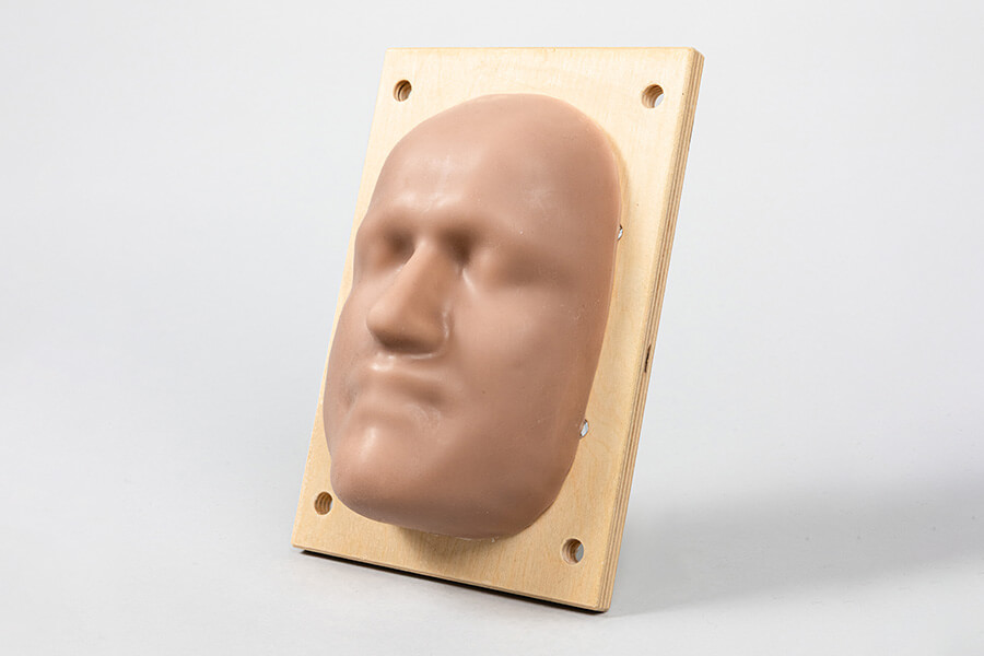
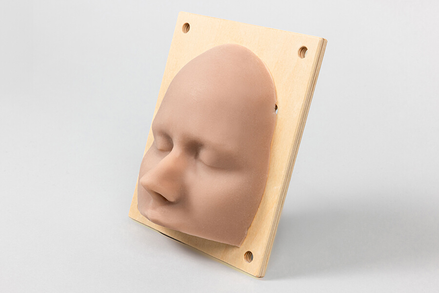
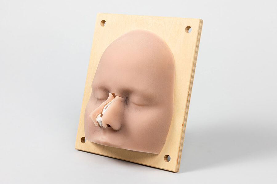
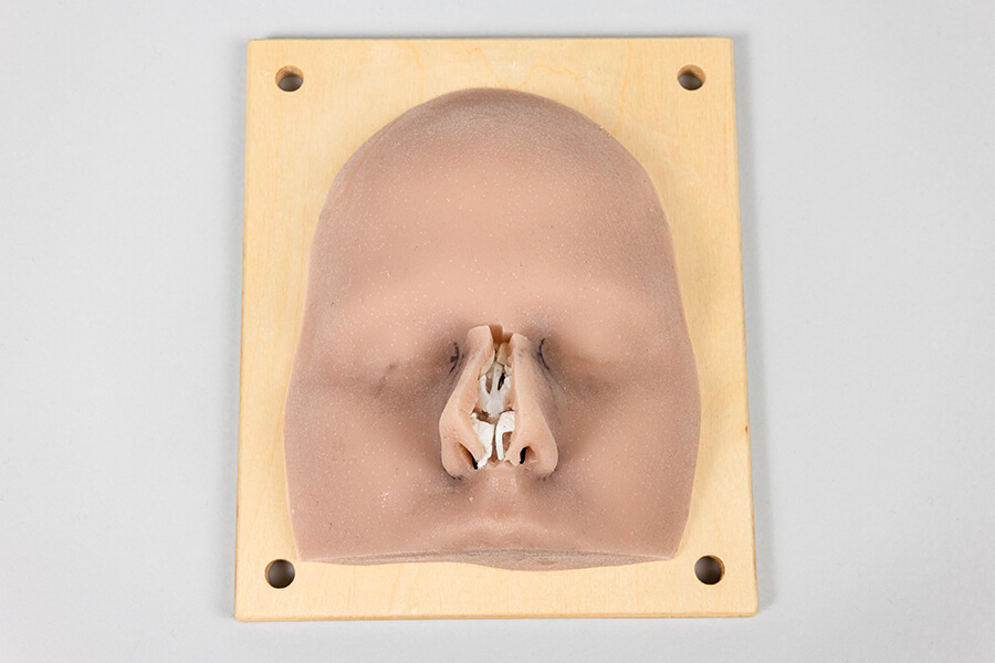
Rhinoplasty model
The model is an artificial replica of the human nose. It consists of a base plate and a silicone layer (5–10 mm) that mimics the thickness of natural human skin. The nose contains artificial nasal cartilage with a transition to the bony area. The base form can be recoated with silicone. Instructions for use are supplied with the model.
Advantages
- Repeatable, uncomplicated practice of various suture techniques
- Simulation possible before operations
- Simplification of various courses and workshops with practical components
Application
- Training for complex operations and reconstructions in the nasal area, e. g.:
- Preparation of the alar cartilage,
- Removal of a bony-cartilaginous hump
- Insertion of a columella graft
- Surgery preparation
- (Specialist) medical training and further education
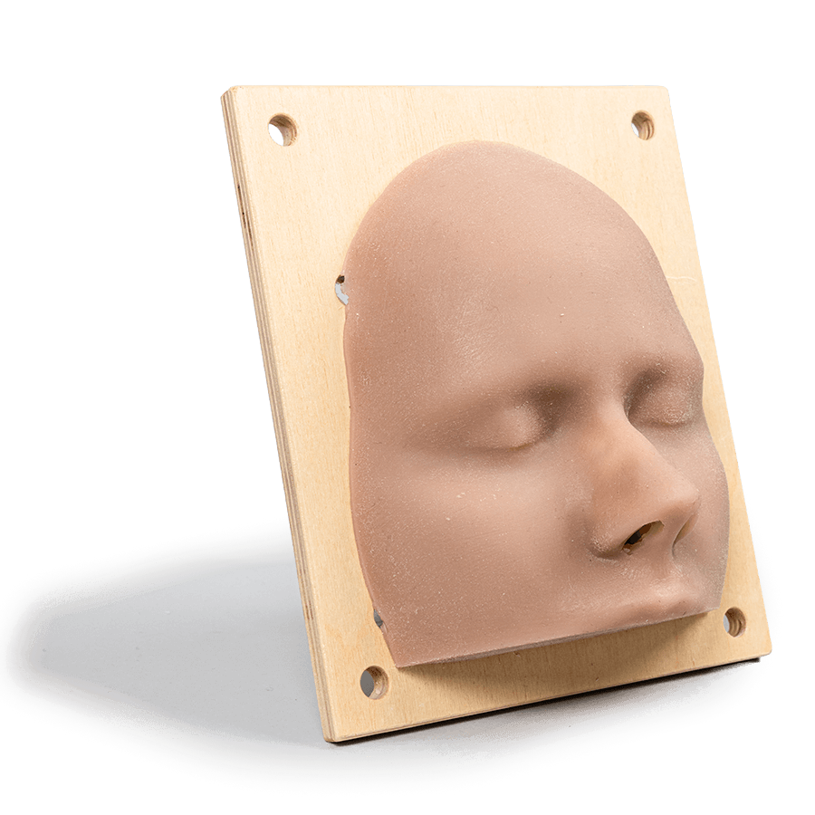
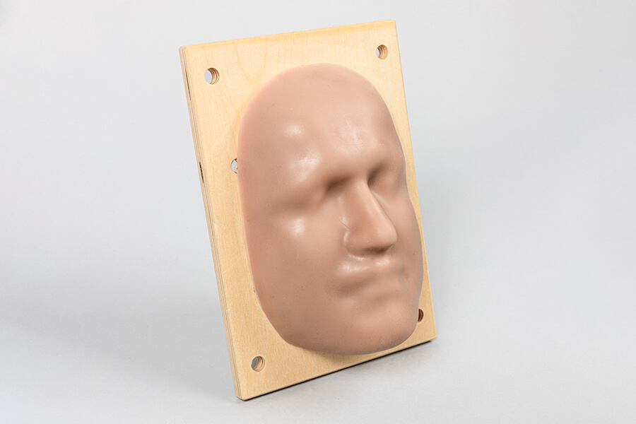
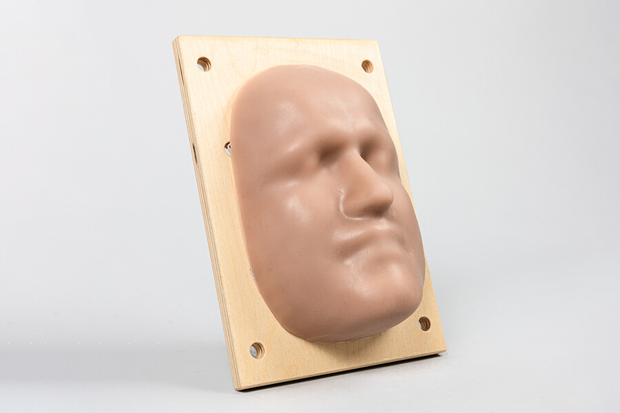
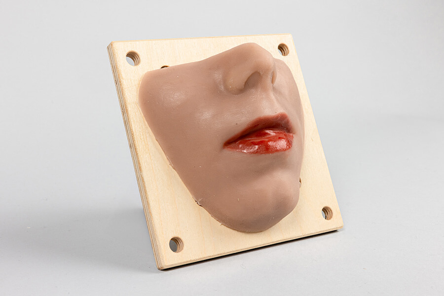
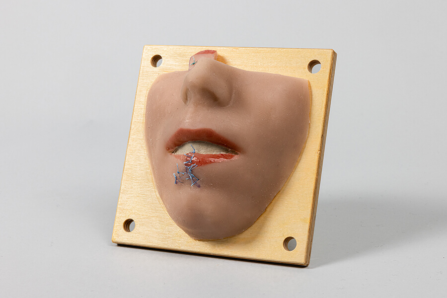
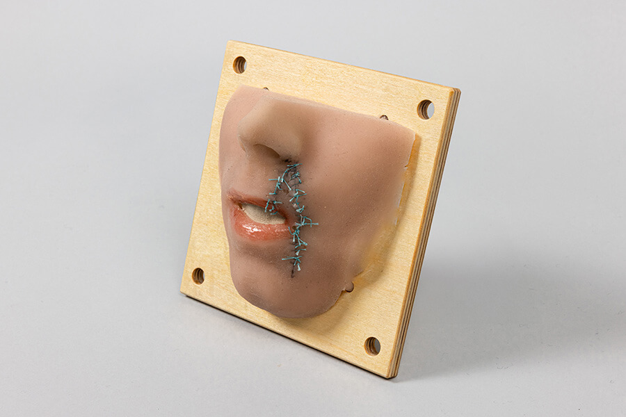
Lip model
The lip model is an artificial replica of the human upper and lower lip and includes a true-to-life representation of the lip-red. The model consists of a base plate and a silicone layer (5–10 mm) that mimics the thickness of natural human skin, with a gauze insert to improve suturability. The base form can be recoated with silicone. The model comes with instructions for use.
Advantages
- Repeatable, uncomplicated practice of various suturing techniques
- Simulation possible before operations
- Simplification of various courses and workshops with a practical component
Application
- Training in complex reconstructions in the lip area, e.g.:
- Defect coverage after tumour resection,
- Flap plasty after tumour resection
- Preparation for surgery
- (Specialist) medical training and further education
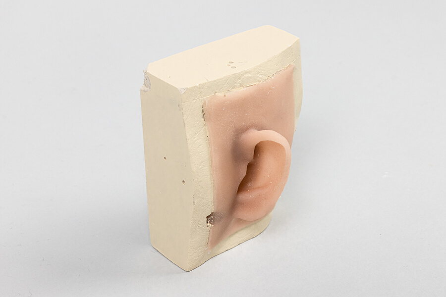
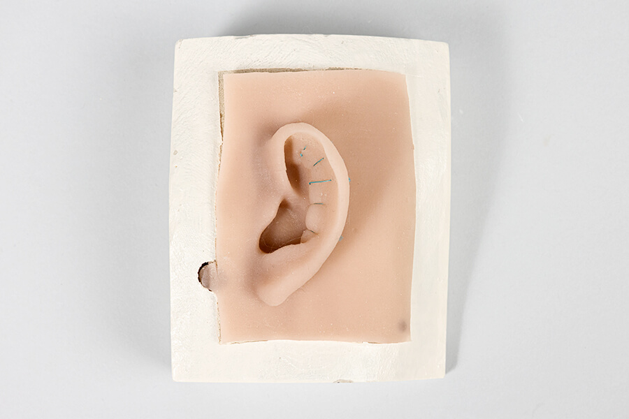
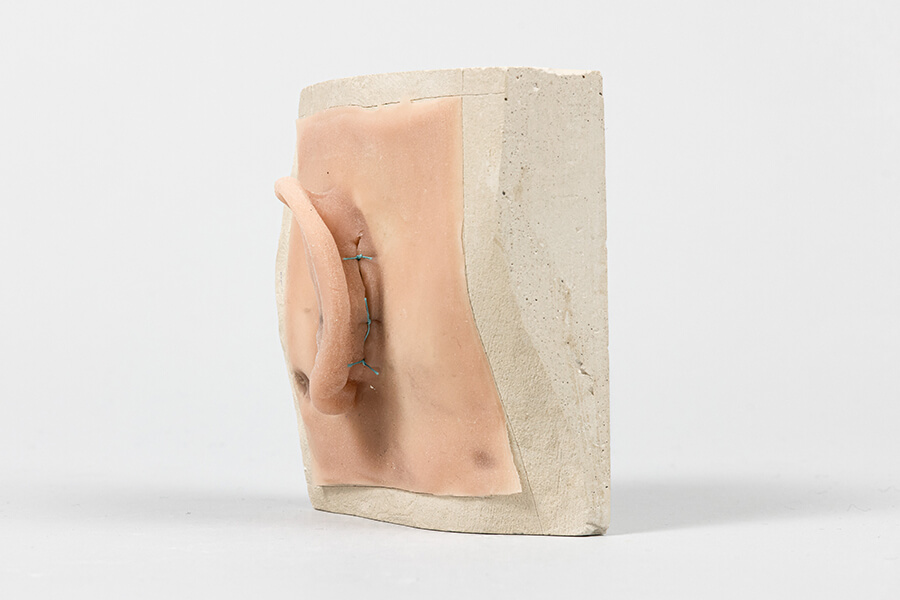
Auricle model
The models consist of a base plate and a silicone layer modelled on the human auricle. The base plate can be recoated.
Advantages
- Repeatable, uncomplicated practice of various suture techniques
- Simulation possible before operations
- Simplification of various courses and workshops with practical components
Application
Antihelix auricle model: artificial replica of grade 1 auricle dysplasia with missing antihelix.
- Training in the formation of the antihelix using pure suture technique
- Training in the refolding of the ear with sutures
- (Specialist) medical training and further education
Cavum auricle model: artificial replica of grade 1 auricle dysplasia with concha hyperplasia.
- Training in cavum resection
- Training in suture techniques for cartilage adaptation
- (Specialist) medical training and further education
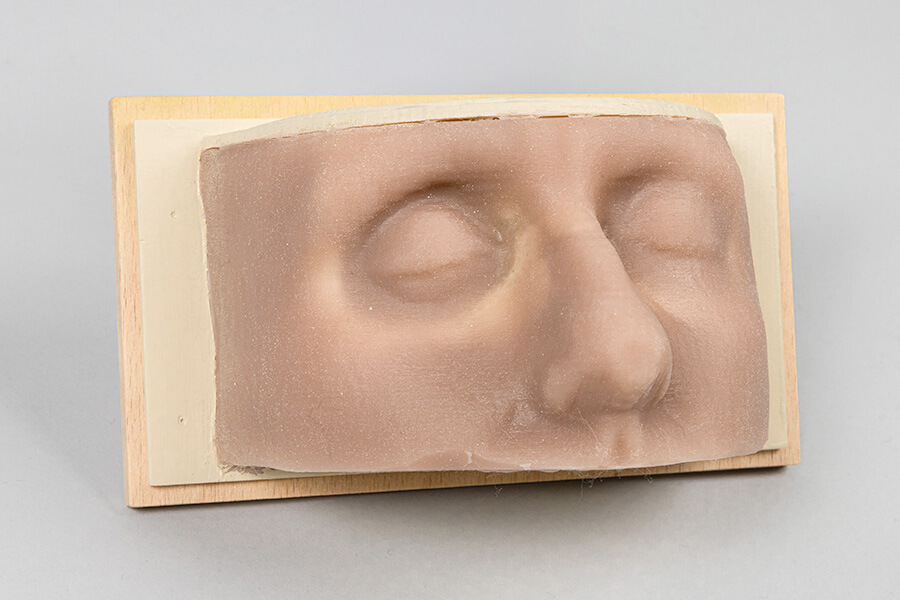
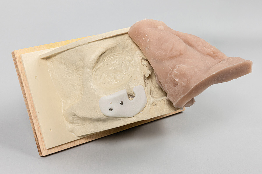
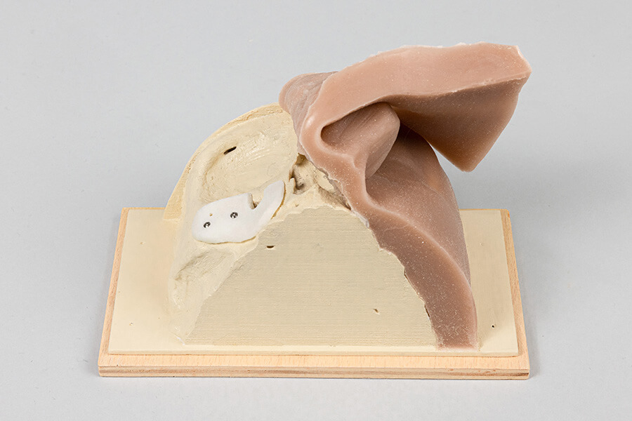
Individual planing model
The individual planning model is a customisable hard and soft tissue model that was developed to simulate the outcome of the operation. The skin surface and bone are represented using real CT data.
Advantages
- Lifelike and detailed simulation and representation of the covering soft tissue in the surgical area
- The models can be edited, separated and the implant model repositioned
- Simplified communication between medical professionals and patients
Application
- Preoperative visualisation of the result (simulation of the patient’s post-operative appearance) in order to prepare for the achievement of an optimal cosmetic result
- Surgical planning in the jaw and orbital area (maxillofacial surgery)
- Practising the correction of malpositions
Do you have any further questions?


